
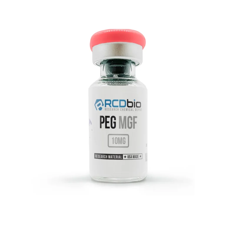

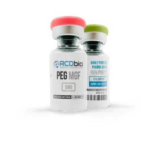

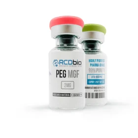
Description
What is PEG-MGF?
PEG-MGF (Polyethylene Glycol Mechano Growth Factor) is a synthetic, PEGylated derivative of Mechano Growth Factor (MGF), the human IGF-1Ec splice isoform of insulin-like growth factor-1 (IGF-1). The native MGF peptide corresponds to the carboxy-terminal Ec domain of the IGF-1 gene, generated through alternative splicing in response to mechanical loading or tissue damage in skeletal muscle. Because the unmodified MGF peptide undergoes rapid proteolytic degradation in biological systems — with a plasma half-life estimated at only a few minutes in preclinical circulation models — the PEGylation modification attaches polyethylene glycol chains to the core peptide, substantially extending its stability and systemic residence time in experimental settings.
The core peptide sequence of PEG-MGF is a 24-amino-acid fragment derived from the Ec extension domain of the IGF-1 gene. Its receptor pharmacology is mechanistically distinct from that of mature IGF-1 and the IGF-1Ea isoform: the MGF E domain has been shown in in vitro systems to act through a receptor pathway that is not blocked by IGF-1 receptor (IGF-1R) antibodies, indicating a receptor interaction independent of the canonical IGF-1R signaling cascade. This distinction makes PEG-MGF a relevant tool compound for investigating paracrine repair mechanisms that differ from systemic IGF-1 signaling.
In research settings, PEG-MGF has been investigated in preclinical models for its role in satellite cell (muscle stem cell) activation, myoblast proliferation, and skeletal muscle regeneration. Additional preclinical research has examined its expression and potential mechanistic activity in cardiac tissue, neural progenitor systems, and bone cell preparations. Peer-reviewed findings across these systems remain limited, and conclusions across experimental models are not always consistent.
Synthetic PEG-MGF supplied by RCDbio is intended strictly for laboratory and research purposes. It is not approved by the Food and Drug Administration for use in this research-grade, non-pharmaceutical form. It is not a dietary supplement and is not intended for human consumption or therapeutic self-administration.
Chemical Properties
| Property | Detail |
| Product Type | Synthetic PEGylated Peptide (IGF-1Ec Splice Variant Derivative) |
| Product Name | PEG-MGF (Polyethylene Glycol Mechano Growth Factor) |
| Application | Scientific / Research Use Only |
| CAS Number | No unique CAS assigned for the PEGylated conjugate; core peptide fragment varies by manufacturer specification |
| Molar Mass | ~2,867.2 g/mol (core peptide, C121H200N42O39); total MW increases with PEG chain size and is vendor-specification dependent |
| Chemical Formula | C121H200N42O39 (core peptide) |
| Sequence | Tyr-Gln-Pro-Pro-Ser-Thr-Asn-Lys-Asn-Thr-Lys-Ser-Gln-Arg-Arg-Lys-Gly-Ser-Thr-Phe-Glu-Glu-Arg-Lys (24-mer Ec domain fragment; PEG conjugation site is N-terminal or lysine-directed per synthesis specification) |
| IUPAC Name | Not formally assigned for the PEGylated conjugate; core peptide follows standard IUPAC amino acid nomenclature |
| Synonyms | Pegylated MGF; PEG-IGF-1Ec; PEGylated Mechano Growth Factor |
| Physical Form | Lyophilized white to off-white powder |
| Solubility | Soluble in sterile water for injection (bacteriostatic water) or acetic acid (0.1–1%); solubility in aqueous buffer may vary with PEG chain length and conjugation site. Gently swirl to dissolve; do not vortex. |
| Storage (Lyophilized) | Store at -20°C in a sealed, light-protected container with desiccant; minimize temperature cycling prior to reconstitution |
| Storage (Reconstituted) | Store at 4°C; use within 48–72 hours of reconstitution. Do not subject it to repeated freeze-thaw cycles. Discard any solution that appears turbid, discolored, or shows particulate matter. |
| PubChem CID | No PubChem CID assigned to the PEGylated conjugate; core Ec peptide fragment data varies by exact sequence specification |
| Purity | ≥98% (HPLC verified, independent third-party laboratory analysis; COA available per batch) |
| WADA Status | PEG-MGF is not listed by name on the current WADA Prohibited List; however, as a growth factor and IGF-1 splice variant, it falls under S2.3 (Growth Factors and Growth Factor Modulators) of the 2026 WADA Prohibited List, which prohibits growth factors affecting muscle, tendon, or ligament protein synthesis. MGF and its derivatives are prohibited at all times in- and out-of-competition. Verify current status at GlobalDRO.com. |
How Does PEG-MGF Work?
PEG-MGF exerts its mechanistic effects primarily through the biological activity of its core Ec domain peptide—the 24-amino-acid carboxy-terminal fragment of the IGF-1Ec splice isoform. The PEG modification does not alter the receptor-interactive peptide sequence; it acts as a steric and hydrodynamic shield that reduces proteolytic access and renal clearance, extending the peptide’s preclinical circulation half-life from minutes to an estimated 48–72 hours in experimental models.
Satellite Cell Activation and Myoblast Proliferation Pathway
In vitro studies using C2C12 myoblast cell preparations have characterized the MGF E domain as a potent activator of satellite cell (muscle stem cell) proliferation. Mechanistically, the Ec domain peptide has been shown to stimulate cell cycle entry in quiescent satellite cells without promoting terminal myogenic differentiation — an effect that is mechanistically distinct from that of mature IGF-1 or the IGF-1Ea isoform. Antibody-mediated blockade of the IGF-1 receptor (IGF-1R) in isolated myoblast preparations did not suppress this proliferative response, indicating that the MGF Ec domain engages a receptor pathway independent of canonical IGF-1R signaling [Yang & Goldspink, 2002]. The identity of this receptor has not been fully characterized in the published preclinical literature.
Mechanical Stimulus-Responsive IGF-1 Gene Splicing
In rodent and human skeletal muscle models, the IGF-1 gene undergoes mechanical load-dependent alternative splicing that transiently upregulates MGF (IGF-1Ec) expression prior to the later expression of systemic IGF-1Ea isoforms. This splicing sequence — MGF expression preceding IGF-1Ea — has been observed in murine in vivo resistance exercise models and in three-dimensional C2C12 cell culture systems subjected to mechanical strain, and has been interpreted preclinically as a temporal regulatory mechanism that prioritizes satellite cell expansion before myotube fusion and hypertrophy [Cheema et al., 2005; Goldspink, 2005].
PI3K/Akt-Associated Downstream Signaling
Preclinical receptor binding and downstream signaling studies have associated MGF E domain activity with activation of intracellular kinase cascades. In murine muscle cell preparations, MGF-mediated signaling has been observed to engage phosphatidylinositol 3-kinase (PI3K) and downstream Akt phosphorylation pathways, which regulate protein synthesis, anti-apoptotic signaling, and cell survival. These observations have been characterized in isolated cell systems and in vivo rodent muscle models; the precise receptor coupling mechanisms upstream of PI3K activation in the context of MGF Ec domain stimulation remain an active area of investigation.
Neuroprotective and Neurogenic Pathway Activity
In transgenic murine overexpression models, MGF has been investigated for activity in neural progenitor cell systems. Overexpression of MGF in mouse brain tissue was associated with increased BrdU-positive proliferating cells in the dentate gyrus and subventricular zone and with preservation of olfactory neuronal function in aging models when overexpression was initiated early [Lalonde et al., 2017]. In the SOD1(G93A) murine model of amyotrophic lateral sclerosis, intramuscular delivery of an MGF expression plasmid was associated with increased motoneuron survival and improved hindlimb muscle strength compared to untreated controls [Riddoch-Contreras et al., 2009]. These findings are specific to gene-delivery and transgenic preclinical systems and do not constitute evidence of activity for the synthetic PEG-MGF peptide in the same model systems.
Key Research Findings
- Myoblast proliferation vs. differentiation: The MGF Ec domain inhibited terminal myogenic differentiation while increasing myoblast proliferation in isolated C2C12 cell preparations; the effect was not blocked by the IGF-1R antibody, suggesting a distinct receptor mechanism. [Yang & Goldspink, 2002]
- Mechanical IGF-1 splicing: IGF-1Ec (MGF) expression was transiently upregulated prior to IGF-1Ea in C2C12 3D cultures under mechanical strain and in rodent resistance exercise models, consistent with a satellite cell expansion role. [Cheema et al., 2005]
- PEG-extended stability: PEGylation of native MGF extended circulation half-life from minutes to an estimated 48–72 hours in experimental preclinical models compared to the unmodified peptide.
- Motoneuron survival: MGF gene delivery in SOD1(G93A) murine ALS models was associated with significantly greater motoneuron survival and hindlimb muscle strength versus controls; effects exceeded those of IGF-1 gene delivery in the same model. [Riddoch-Contreras et al., 2009]
- Hippocampal neurogenesis: MGF overexpression in transgenic murine models increased BrdU-positive proliferative cell counts in the dentate gyrus and olfactory bulbs, associated with preserved olfactory function at 24 months. [Lalonde et al., 2017]
All findings listed above are derived from preclinical or in vitro data. No conclusions regarding human therapeutic efficacy can be drawn from these observations. These findings do not constitute evidence of safety or efficacy in any human condition or organism.
What are the Potential Research Applications of PEG-MGF?
Skeletal Muscle Satellite Cell and Myoblast Biology
PEG-MGF has been employed in preclinical research as a tool compound for investigating satellite cell activation kinetics, myoblast proliferation signaling, and the mechanosensory regulation of the IGF-1 gene. It is suitable for use in in vitro cell culture models — including C2C12 mouse myoblast preparations and primary human myoblast cultures — as well as in vivo rodent muscle injury and overload models, where its extended half-life relative to native MGF permits more stable systemic or local tissue exposure during experimental protocols.
IGF-1 Isoform Pharmacology and Receptor Characterization
Because the MGF Ec domain appears to engage a receptor mechanism distinct from the canonical IGF-1R, PEG-MGF serves as a useful reference compound in studies designed to differentiate IGF-1 isoform-specific signaling from systemic IGF-1 activity. It may be employed in receptor displacement assays, downstream kinase phosphorylation profiling, and comparative growth factor pharmacology experiments involving IGF-1R, insulin receptor, and other RTK systems.
Neurodegenerative Disease Models
Preclinical MGF research in SOD1(G93A) murine ALS models and transgenic neurogenesis models has established a basis for investigating PEG-MGF as a neuroprotective signaling tool. Researchers investigating motoneuron survival, hippocampal neurogenesis, or growth factor-mediated neuroprotection may employ PEG-MGF in gene expression, cell proliferation, or behavioral assays in rodent preclinical systems.
Cardiac and Connective Tissue Repair Research
Preclinical investigations have identified MGF expression in cardiac tissue following myocardial injury and in osteoblast preparations, positioning PEG-MGF as a candidate tool compound for in vitro cardiac cell models and bone cell proliferation assays. These research applications remain at early preclinical stages.
These are observed in preclinical and in vitro contexts only and do not constitute claims of efficacy or safety in any organism.
What are the Potential Side Effects of PEG-MGF?
- Satellite cell over-activation: Supraphysiological concentrations of MGF peptide in isolated muscle cell preparations have been associated with unregulated myoblast proliferation kinetics; dose-dependent effects on differentiation timing have been characterized in C2C12 in vitro systems and may vary across cell types and species
- IGF pathway cross-reactivity: At high concentrations, partial agonism at IGF-1R or insulin receptor systems cannot be excluded based on structural homology with the IGF-1 precursor sequence; observed in cell-based binding assays; not characterized for PEG-MGF specifically at research concentrations
- PEG-related immune responses: High-molecular-weight PEGylated proteins have been associated with anti-PEG antibody generation in rodent and primate in vivo models upon repeated systemic administration; this immune response has not been characterized for PEG-MGF specifically; findings from PEGylated pharmaceutical proteins provide the mechanistic basis for this precaution
- Cardiovascular and mitogenic uncertainty: Preclinical studies on related IGF-1 pathway compounds at supraphysiological concentrations have raised concerns about mitogenic signaling in non-target tissues; specific toxicological data for PEG-MGF at research concentrations in mammalian in vivo models are limited in the peer-reviewed literature
- Neurogenic off-target effects: Preclinical overexpression models showed increased neural progenitor proliferation; the downstream consequences of PEG-MGF exposure on non-target neural tissue at research concentrations have not been fully characterized
No human safety or tolerability data pertaining to research-grade PEG-MGF has been established. These observations are derived from experimental systems and should not be extrapolated to human or animal outcomes.
Risk & Handling
Handling Precautions
PEG-MGF should be handled exclusively by trained laboratory personnel familiar with peptide research compounds. Minimum personal protective equipment includes nitrile gloves, a laboratory coat, and eye protection. Reconstitution of lyophilized powder should be performed in a biosafety cabinet or laminar flow hood to minimize aerosolization and environmental contamination. Avoid generating aerosols during reconstitution. Standard aseptic technique is required for all solution preparation steps. PEG-MGF is not a cytotoxic proapoptotic compound at research concentrations, but its IGF pathway activity warrants appropriate caution when used in proliferating cell systems. Avoid contact with nucleotide-sensitive assay components that may be affected by growth factor contamination.
Exposure Risks
Risk Tier: MODERATE
The biological activity of the MGF Ec domain at the cellular level — specifically its capacity to stimulate satellite cell proliferation and activate PI3K/Akt-associated signaling — classifies PEG-MGF as pharmacologically active at research-relevant concentrations in cell and animal models. Preclinical in vivo toxicity data specific to the PEGylated form are limited in the peer-reviewed literature. The extended half-life conferred by PEGylation (estimated 48–72 hours in circulation versus minutes for native MGF) means that systemic effects in in vivo rodent models may persist considerably longer than with the unmodified peptide. Dose-dependent off-target mitogenic activity at supraphysiological concentrations cannot be excluded based on IGF-1 pathway pharmacology. No human safety data has been established for research-grade PEG-MGF. Researchers should exercise caution appropriate to handling a potent biologically active peptide with growth factor activity.
Storage
- Lyophilized form: Store at −20°C in a sealed, light-protected container with desiccant
- Reconstituted form: Store at 4°C; use within 48–72 hours of reconstitution
- Do not subject to repeated freeze-thaw cycles; peptide integrity and PEG conjugation stability may be compromised with each cycle
- Do not expose to strongly acidic or basic conditions outside the recommended reconstitution buffer range
- Discard any reconstituted solution that appears turbid, discolored, or shows particulate matter
FAQs
Q: What is PEG-MGF, and what is it investigated for in research? A: PEG-MGF (Polyethylene Glycol Mechano Growth Factor) is a synthetic, PEGylated derivative of the MGF Ec domain — the 24-amino-acid carboxy-terminal fragment of the IGF-1Ec splice isoform. In preclinical research, it has been investigated for its role in satellite cell activation, myoblast proliferation signaling, and skeletal muscle repair mechanisms in rodent in vivo models and in vitro cell culture systems. Additional preclinical research has examined its activity in neural progenitor and cardiac cell preparations. It is not approved by the FDA for any human use and is supplied by RCDbio for laboratory and research purposes only.
Q: What is the half-life of PEG-MGF in preclinical models? A: Native MGF (unmodified Ec domain peptide) has a plasma half-life estimated at approximately 5-7 minutes in preclinical circulation models, reflecting rapid proteolytic degradation. PEGylation substantially extends this residence time; the preclinical estimated half-life for PEG-MGF is approximately 48-72 hours in rodent in vivo models, though the precise value may vary with PEG chain molecular weight and conjugation chemistry. These figures are derived from laboratory and preclinical models and do not represent human pharmacokinetic data for research-grade material.
Q: How should PEG-MGF be stored to maintain stability? A: Lyophilized PEG-MGF should be stored at −20°C in a sealed, light-protected container with desiccant until use. Following reconstitution, the solution should be maintained at 4°C and used within 48–72 hours. Repeated freeze-thaw cycles should be avoided, as they may compromise both peptide structural integrity and PEG conjugation stability. Any solution showing turbidity, discoloration, or particulate formation should be discarded.
Q: What reconstitution solvent is recommended for PEG-MGF in laboratory research? A: In laboratory research protocols, PEG-MGF lyophilized powder is most commonly reconstituted in bacteriostatic water (sterile water containing benzyl alcohol as a preservative) or 0.1–1% acetic acid in sterile water, depending on the experimental design and downstream assay requirements. Reconstitution should be performed by gently swirling or rolling the vial; vortexing is not recommended, as mechanical shear stress may disrupt the PEG conjugation or peptide folding. Researchers should consult their specific experimental protocol and downstream assay compatibility requirements before selecting a reconstitution solvent.
Q: How does the MGF Ec domain receptor mechanism differ from that of mature IGF-1? A: In isolated C2C12 myoblast cell preparations, the biological activity of the MGF Ec domain peptide was not blocked by antibody-mediated inhibition of the IGF-1 receptor (IGF-1R), indicating that the Ec domain engages a receptor pathway independent of canonical IGF-1R signaling [Yang & Goldspink, 2002]. Mature IGF-1, by contrast, signals predominantly through IGF-1R-mediated phosphorylation cascades. This mechanistic distinction makes PEG-MGF a useful tool compound for studies designed to separate IGF-1R-dependent from IGF-1R-independent growth factor signaling in skeletal muscle and other tissue systems. The specific receptor through which the MGF Ec domain exerts its proliferative effects in preclinical models has not been definitively identified in the published literature.
Q: What toxicity observations have been reported in preclinical studies involving MGF? A: Compound-specific in vivo toxicity data for PEG-MGF are limited in the peer-reviewed literature as of June 2026. Observations in related preclinical systems include dose-dependent effects on myoblast proliferation kinetics in isolated cell preparations, potential for cross-reactivity with IGF-1R at supraphysiological concentrations, and the general preclinical concern for anti-PEG antibody generation associated with repeated in vivo administration of high-molecular-weight PEGylated compounds in rodent and primate models. Researchers should not extrapolate findings from other PEGylated peptide compounds directly to PEG-MGF without compound-specific verification.
Q: Is PEG-MGF the same compound as native MGF or the unmodified IGF-1Ec fragment? A: No. Native MGF (unmodified Ec domain peptide) and PEG-MGF are structurally and pharmacokinetically distinct. The core 24-amino-acid peptide sequence is the same, but PEG-MGF carries polyethylene glycol chains attached to the peptide backbone — most commonly at the N-terminus or at lysine residue side chains — that substantially alter the compound’s hydrodynamic radius, protease resistance, and systemic half-life. In preclinical research models, native MGF degrades in minutes while PEG-MGF maintains biological activity for approximately 48–72 hours. Experimental protocols designed for one form should not be assumed to apply directly to the other without protocol-specific validation.
Related Research Compounds
IGF-1 LR3 Peptide — A long-arginine extension analog of IGF-1 investigated in preclinical cell and animal models for IGF-1R-mediated anabolic signaling; employed in comparative studies alongside MGF-pathway compounds to differentiate receptor-specific growth factor responses.
BPC-157 Peptide — A synthetic pentadecapeptide investigated in rodent in vivo models for tissue repair and cytoprotective signaling; commonly used in preclinical regenerative biology research alongside growth factor peptides.
GDF-8 (Myostatin) Peptide — A TGF-β superfamily member investigated as a negative regulator of skeletal muscle mass in preclinical models; relevant to comparative studies examining anabolic and anti-anabolic growth factor signaling in muscle biology research.
All products listed are for laboratory and research purposes only.
References
- Yang SY, Goldspink G. (2002). Different roles of the IGF-I Ec peptide (MGF) and mature IGF-I in myoblast proliferation and differentiation. FEBS Letters, 522(1-3):156–160. https://pubmed.ncbi.nlm.nih.gov/12095637/
- Goldspink G. (2005). Mechanical signals, IGF-I gene splicing, and muscle adaptation. Physiology, 20(4):232–238. https://pubmed.ncbi.nlm.nih.gov/16024511/
- Cheema U, Brown R, Mudera V, Yang SY, McGrouther G, Goldspink G. (2005). Mechanical signals and IGF-I gene splicing in vitro in relation to development of skeletal muscle. Journal of Cellular Physiology, 202(1):67–75. https://pubmed.ncbi.nlm.nih.gov/15389530/
- Riddoch-Contreras J, Yang SY, Dick JRT, Goldspink G, Greensmith L, Orrell RW. (2009). Mechano-growth factor, an IGF-I splice variant, rescues motoneurons and improves muscle function in SOD1(G93A) mice. Experimental Neurology, 215(2):281–289. https://pubmed.ncbi.nlm.nih.gov/19038252/
- Lalonde R, Strazielle C. (2017). Mechano-growth factor promotes axonal sprouting and increases the number of olfactory receptor neurons in aging mice. Molecular Brain, 10(1):39. https://pubmed.ncbi.nlm.nih.gov/28683812/
Disclaimer
PEG-MGF is exclusively for laboratory research purposes. RCDbio products are not intended to diagnose, prevent, treat, or cure any disease or medical condition.
The Food and Drug Administration has not evaluated the statements on our website. This product is not approved for human or veterinary use. Researchers must comply with all applicable local, state, and federal laws and regulations governing the purchase and use of research compounds. By purchasing, you agree to our Terms and Conditions. RCDbio reserves the right to refuse sales to unauthorized individuals.
ATTENTION: All RCDbio products are strictly for LABORATORY AND RESEARCH PURPOSES ONLY. They are not intended for human consumption, veterinary use, or any other non-research application. For queries, complaints, or support, contact support@legacy.rcdbio.co
Reviews
There are no reviews yet